Female Upper Thigh Anatomy - There are two hip bones, one on the left side of the body and the other on the right.. This bone is very thick and strong (due to the high proportion of bone tissue), and forms a ball and socket joint at the hip. The largest of them is the most superficial muscle, the gluteus maximus. More images for female upper thigh anatomy » The thigh muscles are divided into three compartments: It helps maintain erect posture, abducts the thigh, and rotates the thigh outward.
The largest of them is the most superficial muscle, the gluteus maximus. Sometimes obturator externus muscle is also considered as part of the hip adductors since one of its actions is to adduct the thigh at the hip joint. The thigh muscles are divided into three compartments: People who play soccer have these specific muscles of the leg very well defined, so they're like a walking anatomy atlas for thigh muscles. Human muscles · july 20, 2016.
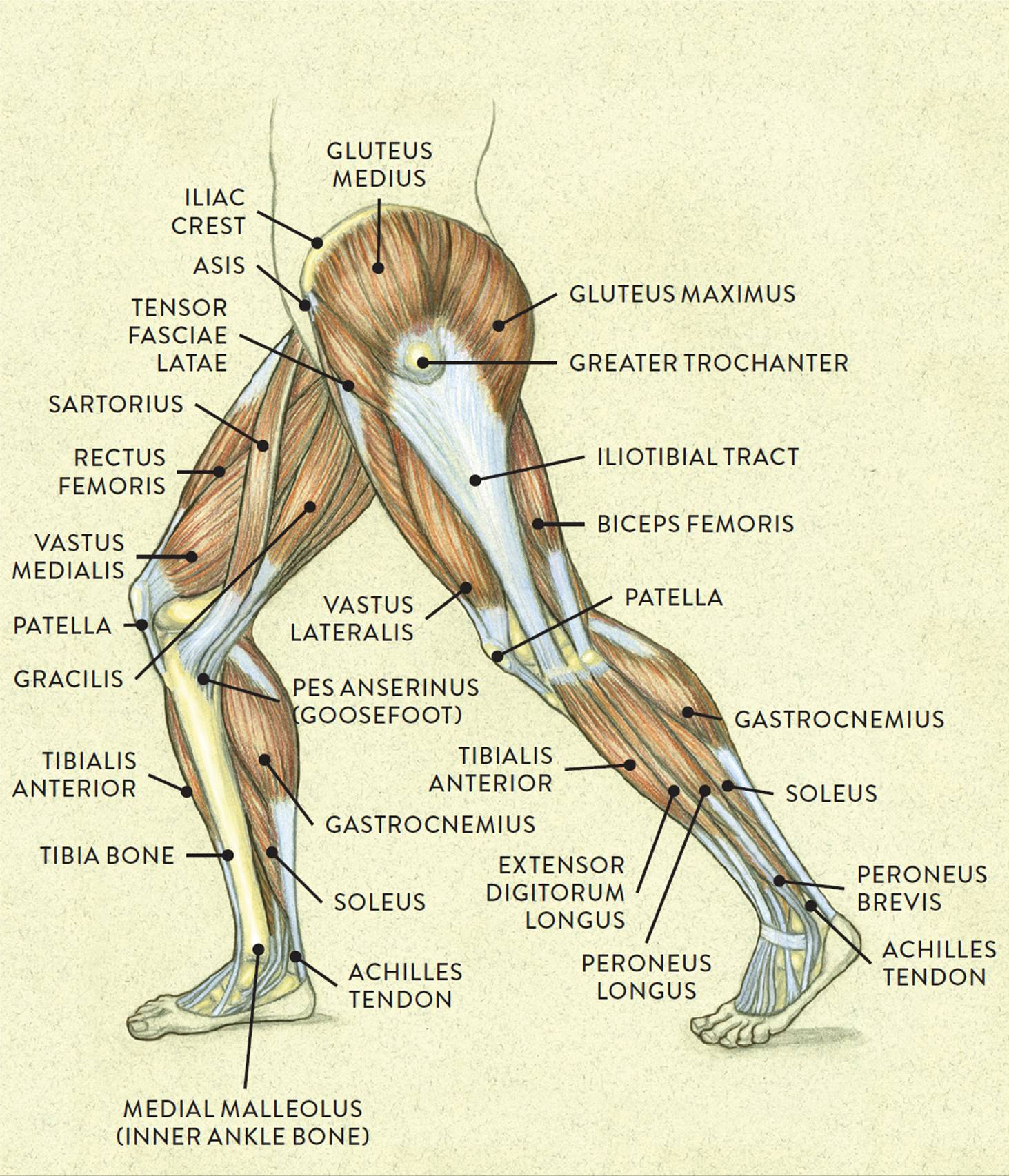
Muscles Of The Leg And Foot Classic Human Anatomy In Motion The Artist S Guide To The Dynamics Of Figure Drawing from doctorlib.info See more ideas about female bodies, anatomy, female anatomy. Vastus lateralis vastus medialis vastus intermedius rectus femoris Sometimes obturator externus muscle is also considered as part of the hip adductors since one of its actions is to adduct the thigh at the hip joint. There are two hip bones, one on the left side of the body and the other on the right. The adductors all originate from the lower pubic bone on the pelvis and insert all along the inner surface of the femur. Below the gluteus maximus is the smaller gluteus medius. Doctor, scientist, specialist in anatomy indicates pointer of obturator foramen where canalis obturatorius, involving obturator ar. People who play soccer have these specific muscles of the leg very well defined, so they're like a walking anatomy atlas for thigh muscles.
The largest of them is the most superficial muscle, the gluteus maximus.
Tery, vein and nerve and. The six hip adductor muscles are all located in the adductor or medial compartment of the thigh and all mainly adduct the thigh at the hip joint. See more ideas about female bodies, anatomy, female anatomy. Lunges are a terrific exercise for strengthening the gluteal muscles, and also the upper thighs. The thigh is the region between the hip and knee joints. These are gracilis, pectineus, adductor longus, adductor brevis, adductor magnus, and adductor minimus muscles. Females typically have a wider pelvis than males to allow for childbirth and this causes the femur or thigh bones to also be positioned wider. Anatomy of human knee joint. People who play soccer have these specific muscles of the leg very well defined, so they're like a walking anatomy atlas for thigh muscles. The thigh muscles are divided into three compartments: This bone is very thick and strong (due to the high proportion of bone tissue), and forms a ball and socket joint at the hip. There are two hip bones, one on the left side of the body and the other on the right. Doctor, scientist, specialist in anatomy indicates pointer of obturator foramen where canalis obturatorius, involving obturator ar.
Adductor magnus, adductor longus, adductor brevis, pectineus, and gracilis. Together, they form the part of the pelvis called the pelvic girdle. Lunges are a terrific exercise for strengthening the gluteal muscles, and also the upper thighs. See more ideas about female bodies, anatomy, female anatomy. These are gracilis, pectineus, adductor longus, adductor brevis, adductor magnus, and adductor minimus muscles.
Shutterstock Puzzlepix from image.shutterstock.com Branches of left subclavian artery. See more ideas about female bodies, anatomy, female anatomy. The adductors are a complex of five muscles which adduct the thigh (pull the thigh inward toward the midline of the body): Human muscles · july 20, 2016. Human knee anatomy with femur, tibia and fibula bones isolated on black. Muscle anatomy of upper thigh. The thigh muscles are divided into three compartments: Below the gluteus maximus is the smaller gluteus medius.
The six hip adductor muscles are all located in the adductor or medial compartment of the thigh and all mainly adduct the thigh at the hip joint.
Vastus lateralis vastus medialis vastus intermedius rectus femoris Tery, vein and nerve and. Lunges are a terrific exercise for strengthening the gluteal muscles, and also the upper thighs. Females typically have a wider pelvis than males to allow for childbirth and this causes the femur or thigh bones to also be positioned wider. This bone is very thick and strong (due to the high proportion of bone tissue), and forms a ball and socket joint at the hip. More images for female upper thigh anatomy » The largest of them is the most superficial muscle, the gluteus maximus. People who play soccer have these specific muscles of the leg very well defined, so they're like a walking anatomy atlas for thigh muscles. Muscle anatomy of upper thigh. Together, they form the part of the pelvis called the pelvic girdle. Below the gluteus maximus is the smaller gluteus medius. Anatomy of human knee joint. There are two hip bones, one on the left side of the body and the other on the right.
More images for female upper thigh anatomy » Below the gluteus maximus is the smaller gluteus medius. Sometimes obturator externus muscle is also considered as part of the hip adductors since one of its actions is to adduct the thigh at the hip joint. The adductors all originate from the lower pubic bone on the pelvis and insert all along the inner surface of the femur. Lunges are a terrific exercise for strengthening the gluteal muscles, and also the upper thighs.
Inner Thigh Muscle Anatomy Anatomy Drawing Diagram from i.pinimg.com Human knee anatomy with femur, tibia and fibula bones isolated on black. See more ideas about female bodies, anatomy, female anatomy. The thigh is the region between the hip and knee joints. These are gracilis, pectineus, adductor longus, adductor brevis, adductor magnus, and adductor minimus muscles. The adductors all originate from the lower pubic bone on the pelvis and insert all along the inner surface of the femur. Tery, vein and nerve and. Vascular anatomy of the upper arm. Human muscles · july 20, 2016.
See more ideas about female bodies, anatomy, female anatomy.
Doctor, scientist, specialist in anatomy indicates pointer of obturator foramen where canalis obturatorius, involving obturator ar. Vastus lateralis vastus medialis vastus intermedius rectus femoris Tery, vein and nerve and. It helps maintain erect posture, abducts the thigh, and rotates the thigh outward. The six hip adductor muscles are all located in the adductor or medial compartment of the thigh and all mainly adduct the thigh at the hip joint. Lunges are a terrific exercise for strengthening the gluteal muscles, and also the upper thighs. People who play soccer have these specific muscles of the leg very well defined, so they're like a walking anatomy atlas for thigh muscles. More images for female upper thigh anatomy » The thigh muscles are divided into three compartments: Vascular anatomy of the upper arm. Human muscles · july 20, 2016. Muscle anatomy of upper thigh. Adductor magnus, adductor longus, adductor brevis, pectineus, and gracilis.
It helps maintain erect posture, abducts the thigh, and rotates the thigh outward upper thigh anatomy. These are gracilis, pectineus, adductor longus, adductor brevis, adductor magnus, and adductor minimus muscles.


Posting Komentar
0 Komentar